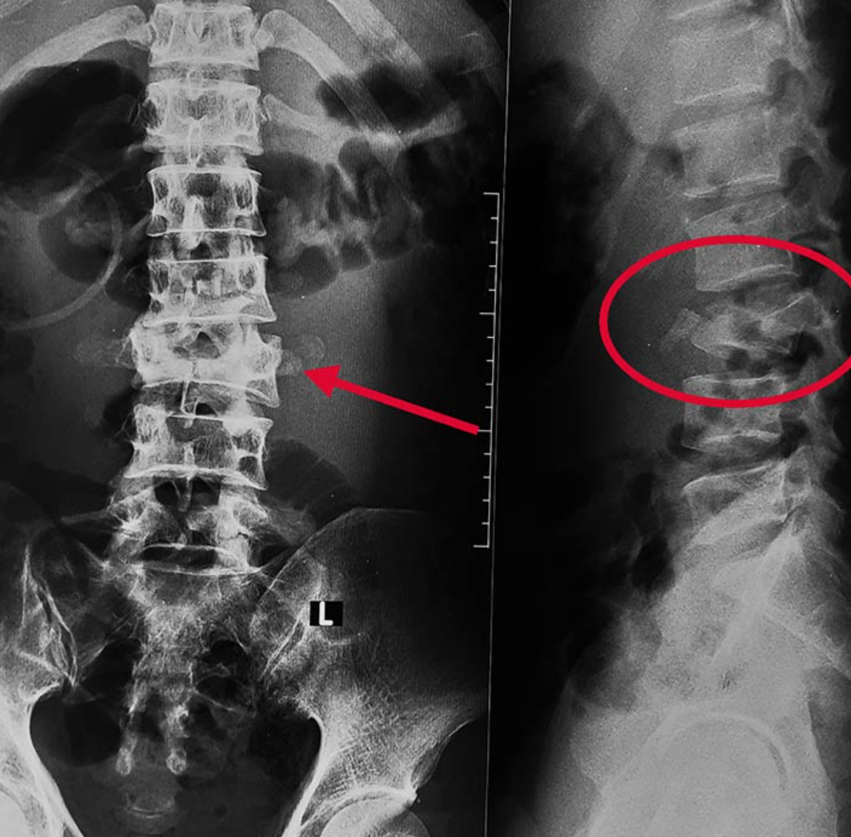
Magnetic Resonance Imaging, commonly known as MRI, is a powerful tool in diagnosing and monitoring the condition of a herniated disc. An MRI provides clear and detailed images of the body’s internal structures, which can be invaluable in understanding the severity and exact location of a herniated disc. In this blog post, we will delve into the role and utility of MRI in diagnosing and evaluating herniated discs.
Role of MRI in Diagnosing Herniated Discs
If you’re experiencing symptoms like back pain, numbness, or weakness, your healthcare provider may order an MRI to help determine the cause. In the case of a suspected herniated disc, an MRI can confirm the diagnosis and provide essential information on the location and severity of the herniation.
What Does a Herniated Disc Look Like on MRI?
A typical MRI image of a healthy spine shows the spinal discs as white structures sandwiched between the darker vertebral bodies. When a disc is herniated, the MRI may reveal an outpouching or displacement of the disc material, typically appearing darker than the rest of the disc. The location of this displacement can help identify the specific disc that’s herniated, such as L4-L5 or L5-S1, which are among the most commonly affected areas.
Understanding MRI Views: Axial vs. Sagittal
When looking at an MRI report or images, you may come across the terms ‘axial view’ and ‘sagittal view.’ These refer to the direction of the image slices. The axial view is like looking at the body from the top down or bottom up, while the sagittal view is a side-on image, like looking at a person’s profile. Both views can provide valuable insight into the condition of the spine and discs.
Reading an MRI Report
An MRI report typically includes a detailed description of the observed structures and any abnormalities. If a herniated disc is present, the report may mention the location of the herniation (e.g., L4-L5 or L5-S1), the size or severity of the herniation, and whether it is compressing any nearby structures like spinal nerves.
However, it’s important to discuss the results with a healthcare professional, as the clinical significance of the findings may depend on various factors, including your symptoms and overall health.
The Process of Getting an MRI
If you’re scheduled for an MRI for a herniated disc, here’s what you can expect: You’ll be asked to lie on a movable table that slides into the MRI machine. The procedure is painless, but it can be a bit noisy due to the machine’s operation. Depending on the area being scanned and the specifics of the study, the procedure may take between 30 minutes to an hour.
In Conclusion
MRI plays an essential role in diagnosing, evaluating, and monitoring herniated discs. It offers a non-invasive and highly detailed look at the spine, helping healthcare professionals devise the most effective treatment plan for their patients. As always, it’s crucial to discuss any concerns or questions about your condition or MRI results with your healthcare provider, as they are best equipped to interpret these in the context of your overall health and symptoms. Understanding the process and findings of an MRI can help you become a more informed and proactive participant in your healthcare journey.
Chiropractic Treatment for Herniated Discs
The approach of chiropractic treatment for herniated discs is typically holistic and patient-centered, aiming to provide relief without surgical intervention or drugs.
Spinal Manipulation: This is the most well-known chiropractic technique, also called chiropractic adjustment. The chiropractor applies a controlled, sudden force to the affected spinal joint, which may help to align the spine, improve spinal function, and alleviate pain. This technique, when done correctly by a professional, is considered safe but should not be performed if certain contraindications exist, like severe osteoporosis or spinal cancer.
Flexion-Distraction Technique: This is a gentle, non-thrust type of spinal manipulation often used for herniated disc treatment. The chiropractor uses a special table that distracts or stretches the spine, and by using a pumping action on the disc instead of direct force, the pressure on the disc can be reduced, leading to pain relief.
Pelvic Blocking Techniques: These methods involve placing cushioned wedges on the sides of the patient’s pelvis, along with gentle stretches. This can help move the disc away from the nerve, reducing inflammation and pain.
Physical Therapy Modalities: Many chiropractors also employ physical therapy techniques, such as ice and heat therapy, electrical stimulation, or ultrasound to reduce inflammation and muscle spasm associated with herniated discs.
Exercises and Lifestyle Modifications: A chiropractor can provide exercises tailored to the patient’s condition, which can help strengthen the spinal muscles and prevent further injury. They may also offer advice on posture and ergonomics to help prevent disc herniation in the future.


